
The absurd amount on sharpening stones on the market should ring some alarm bells. The first is: there must be a lot of money in this. The second: what’s the difference between them? The third: which is the ideal one (for me)?
I’ve ordered and then analysed a couple of different grinding stones. This is probably going to become an ongoing series of blog posts, whenever I get new and exciting grinding stones. If you have some suggestion on what I should look at next, or want to share your super secret DIY stones, I could be persuaded to open the bag of analytical devices… hit me up on Instagram under @marvgro for that.
Fällkniven DC3 (diamond/ceramic whetstone)
According to the manufacturer’s homepage, this is a “diamond grit 25 micron, sapphire ceramic grit 5 micron”. Let’s take a look!


Optical Micrograph of the diamond side. Magnification and scale bar are visible on the lower right part of the image. Microscope: Leica Emspira
The diamond side is coated in TiN. Typically, this coating can be found on cheaper HSS tooling, as it’s quite hard (2400-2700 HV), but also slick and doesn’t let chips adhere. It’s a curious choice to put on a grinding stone, as the grit used here (diamond) is quite a bit harder – depending on the grain orientation, it clocks in at 10000 HV. It’s certainly nice looking though, and I’d postulate that this is the main reason it is applied to any sharpening stone.


SEM Micrographs of the Fällkniven DC3 stone. It shows quite a large range of grain sizes. Instrument: Thermo Fischer PhenomXL Scanning Electron Microscope
SEM pictures show gritty, sharp diamonds. The range distribution of the visible grains (measured at their largest diagonal distance) ranges from 50 to 75 micrometre, with a strong weighting towards the upper end. The grit’s have a distinct checkered look to them – this is the coating, sticking to some parts of the diamond, and not adhering to others. It is very likely that the first use of the stone would remove the coating at any point that is in contact with a blade.


Energy dispersive x-ray spectroscopy (EDS) inside the scanning electron microscope show the coating (Ti, N), the diamond grain (C) as well as the galvanic binder around the grains (Ni). Instrument: Thermo Fischer PhenomXL Scanning Electron Microscope
In order to faciliate a better sense of depth and size, a surface scan was undertaken via white light interferometry. This creates a very high resolution height map – the Z resolution here is absurdly small, where’s the X/Y resolution (“spatial resolution”) follows the Abbe diffraction limited law.

White light interferometry height map of the diamond surface. Instrument used: Zygo Nexview NX2, Objective Lens: 10X. Stitched overview of 4×4 images.
We can see the typical galvanic bound height distribution – unevenly spaced grains with some very high outliers. This is the main reason that galvanic stones leave larger scratches and commonly a worse surface than a similar grain sized vitrified or resin bound stone.

ISO 25178 surface parameters of the Fällknives DC3 diamond side.
The ISO25178 parameters show a rough surface (Sa/Sq are the arithmetic respective quadratic surface roughness). Sz is the total height of the surface. Very indicative of the distribution is the parameter “Sdc”, which shows the range between the lowest 10% and highest 90% of the measured points. This is a good indicator how “even” the height distribution is. A perfect flat surface would have a value of 0 here, whereas a widely spread surface shows a wider range. It’s a usefull parameter to compare stones, but leaves out the 10% outliers at every end. Sku, the kurtosis shows how “sharp” the surface data is. Typically, a value below 3 is considered flat, whereas values above 3 are considered very sharp.
The other “ceramic” side shows a typical ceramic abrasive mix.


SEM images show a pretty uniform, surface with some large voids.
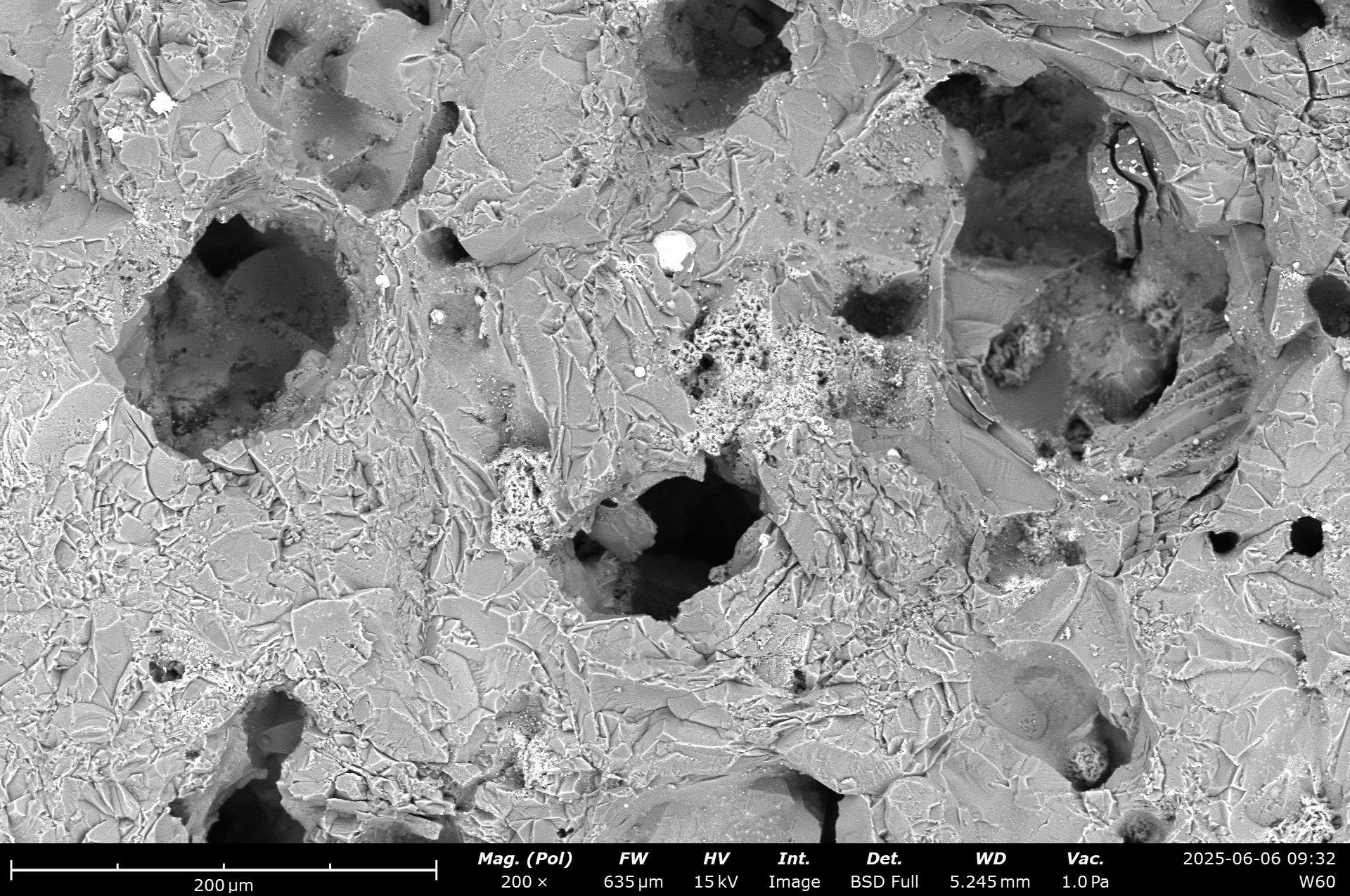


SEM micrographs of the surface morphology. A typical, sintered alumina oxide appearance with some foreign particles (darker colour in the BSD image) and large voids are visible. Instrument: Thermo Fischer PhenomXL Scanning Electron Microscope
While the void size is suprising, this certainly allows for some swarf build-up. 🙂 Some metal particles (bright white colour), but also some different abrasive grains (slightly darker grains) are visible. The detector used is a back-scatter detector. Here, besides the topographical contrast, one also has a contrast based on the weight of the element. The rule of thumb here is: the heavier the element, the brighter the returned pixel is. Pure metals are typically the brightest, whereas ceramics or diamonds are of darker colour.

EDS analysis of the chemical composition. The colour corresponds to the individual element, visible above the scale bar. Instrument: Thermo Fischer PhenomXL Scanning Electron Microscope
Chemical analysis show several large SiC grains, as well as Al2O3 grains. As sapphire is chemically Al2O3, just in a monocrystalline configuration, I think we have identified plenty about the compoistsion. Trace elements of metals and Calciumoxide (blue colours) are likely impurities from manufacturing.

White light interferometry height map of the ceramic surface. Instrument used: Zygo Nexview NX2, Objective Lens: 10X. Stitched overview of 4×4 images.
The whitelight interferometry surface map shows a relatively rough surface. large voids are visible, the range of height values doubles compared to the diamond size. On the other hand, the uppermost part of the surface shows a higher plateau region. The contact area likely is higher on this stone side. Sku, the kurtosis shows how “sharp” the surface data is. Typically, a value below 3 is considered flat, whereas values above 3 are considered very sharp. Here, a much lower value than on the diamond surface can be seen.
Combined with the low sharpness of the dull ceramics, a burnishing effect is expected, improving the appearance of a blade with very low effort.

ISO 25178 surface parameters of the Fällknives DC3 ceramic side.
Leave a Reply