

This is part of a series of blog posts – looking into the appearance and composition of commercially available sharpening stones. If you are interested in the previous episodes, check out the archive for them.
If you have some suggestion on what I should look at next, or want to share your super secret DIY stones, I could be persuaded to open the bag of analytical devices… hit me up on Instagram under @marvgro for that.
Disclaimer: I’m not for sale. Every review you see on this blog is bought with my own money. I have no affiliation to any manufacturer.
Review
Today’s stone is a very famous, well known brand. Naniwa is a Japanese brand, that has been making sharpening stones for over 60 years. Their homepage says they deal in all things abrasive. I like that! The stone I have bought is from the chosera line, with 5000 grit. It is, according to diverse homepages, an alumina-oxide stone with a magnesia binder. Let’s take a closer look:


Optical micrograph of the Naniwa chosera 5000. Instrument: Leica Emspira.
It has some marbeling to it, with a fine and smooth surface. Zooming in, individual particles and grains become visible. For a closer look, as usual, we take a look in the SEM!


SEM micrographs of the Naniwa Chosera 5000 stone. Instrument: Zeiss GeminiSEM560.
At low magnifications, the stone appears to have a smooth cover above the abrasive grit, covering about 80% of the surface. Zooming in further, one starts to identify cubic, small abrasive grits, but also that the covering “film” actually consists out of an uncountable amount of sub-µm particles, that are slightly rounded and longish. This is an interesting stone! To identify this, we will employ EDS analysis – check out this segment of the blog to understand the SEM metrology better. I mentioned before, that things get… tight once you employ the full suit of sensors. Because this stone is non-conductive, but we need a lot of acceleration voltage to get a reliable EDS reading, I’ve employed our low vacuum mode. With the aid of a small orfice, it is possible to increase the chamber pressure, but leave all sensors functional. For this, a diode-type BSD sensor with the small aperture is inserted pneumatically into the chamber. The EDS sensor meanwhile is shaped like a pen, coming in from the other side. To show you how unbelievably tight and confusing everything gets, I snapped you a shot of the chamberscope:

View from the chamberscope. The stone is visible diagonally from lower left to upper right. The EDS sensor is the pen shaped object coming from the right upper corner. The low-vacuum aperture sits below the pyramidal pole piece, and has been inserted from the left side of the picture. Instrument: Zeiss GeminiSEM560.
The EDS analysis reveals the chemical composition:


EDS analysis of the Naniwa Chosera 5000. Instrument: Oxford Ultim Max ∞ 40mm2 EDS sensor. Note that our EDS sensor doesn’t show elements lighter than boron.
There’s a fascinating mix of chemical elements up there. The old truth “you can find the whole periodic table in ceramics” hold’s especially true. Very interesting is the large particle, with flaky composition. Let’s zoom in a bit more on that one:

SEM micrograph of a flaky particle, found in the Naniwa Chosera 5000 stone. Instrument: Zeiss GeminiSEM 560.

Elemental analysis of the flaky particle. Instrument: Oxford Ultim Max ∞ 40mm2 EDS sensor. Note that our EDS sensor doesn’t show elements lighter than boron.
This is really fascinating! we can pick out what we expected – Mg, O, Al, but also massive amounts of silicon. This is the moment where I am really happy, I haven’t spend more time studying minerals, because I don’t even want to imagine what this could be. Nevertheless, I went on a literature deep dive for you folks. There’s a couple of possibilities this could be – a paper I found had something similar to the flakes we are seeing, but was writen by geologists1. They stated that the flakes they are seeing could either be: Anortit – CaAl2Si2O8; Albit – NaAlSi3O8; and Paligorskit – (Mg,Al)5(Si,
Al)8O20(OH)28H2O. The nice thing about geology is, pretty much every mineral has decent SEM pictures online. Albit2 has a matching appearance3, but the excited reader might now ask, where is our Sodium (Na) at this point? Well, it’s pretty exchangeable with Calcium (Ca), which we found in our spectrum. I have no idea what this is called, and I am quite unsure at this point what we found. Structure wise, it should be a triclin crystal latice, and it consists out of the chemical elements Si, Mg, O, Ca. If anyone has studied geology and wants to supply the solution here, reach out. If not, I now declare this to be something like Albit. It doesn’t really matter – it just shows they sinter these stones at quite the high temperature, and there’s cool structures hidden in the microcosmos! All of these oxides are of similar hardness – quite a bit harder than steel, not much harder than carbides.
Let’s take a look at the surface composition!

Instrument: Bruker Alicona µCMM, 50X objective lens, 3×3 FOV high resolution focus variation scan. Data is leveled and outliers removed (0.25%).
It’s a smooth stone, without a large amount of bearing surface. Roughness is not exceptionally smooth, but there are also no super deep recesses. This is a finely made stone, and if you scratch along the surface with your fingernail, there’s nothing catching it. The feedback, because of the relatively rough surface is noticeable. This stone doesn’t glitch over your knife edge.

ISO 25178 parameters of the Naniwa Chosera 5000 stone.
In order to evaluate the sharpening performance of this stone, a blade was sharpened with it. I am using a standardised testing procedure, read about it here. Nevertheless, it’s 65 HRC M398, and sharpened to 17 DPS with resin bond diamond stones down to 10 µm. Afterwards, the tested stone is used, first in a back and forth movement until the surface becomes homogenous, and then alternating strokes (5-5-3-2) on each side, for a total of 20 strokes towards the apex per side. No pressure is applied but the weight of the apparatus. The stone was only splashed with water, not soaked (in accordance with the website I bought it from).


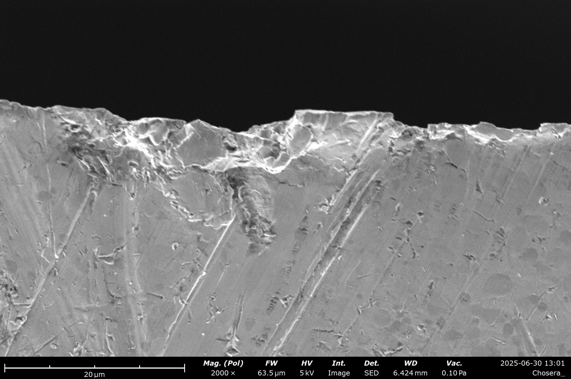
SEM micrographs of the blade finished with the Naniwa Chosera. A slight cross hatch pattern is visible, which stems from me changing hands during the sharpening. The edge is burnished at some parts, whereas other parts show fractures. Overall, the appearance diminished a bit compared to the 10 µm resin stone.
The edge shows no burr, but some cracking near the carbides, brittle failure of the edge and larger scratches are visible. I think the stone struggled a lot with the very hard (65 HRC!) high carbide steel, but also the fact that on a guided system, you can only splash it with water – it doesn’t build up a slurry. Seriously? I think this might be a fantastic stone for freehand sharpening and lower hardness steels. On this guided system, with the minimum amount of water I was able to apply, I am not a super large fan.
I will try to revisit this stone in the future with a softer steel.
References:
- Paper: Cvetković, Vesna & Purenović, M.M. & Jovićević, Jovan. (2006). Change of water electrochemical characteristics in contact with magnesium enriched kaolinite-bentonite catalyst substrate. CHISA 2006 – 17th International Congress of Chemical and Process Engineering. ↩︎
- https://en.wikipedia.org/wiki/Albite ↩︎
- https://www.google.com/search?sca_esv=6d27a280cc9bfc7a&rlz=1C5CHFA_enDE1085DE1085&sxsrf=AE3TifN5R8VHVx9-SUBmyiUP492AV-8O9w:1751306917124&udm=2&fbs=AIIjpHw2KGh6wpocn18KLjPMw8n5Yp8-1M0n6BD6JoVBP_K3fa3EquCb45pN-svRz-qicbUuJAUxGdF5oHE4vP7-4OPt4Q9Fw9x1rJO_JqPfqqyI0sgiH2ECQfCuqfNq42mWqHj89AzOZLwcp1D39M5NMYTfJhenwM2DIDoriG9lJQRIvRH6btwwjWjRRvECqtMI6DdjJ1FlpC6XGLXqienEXDu0DUd7Ig&q=albit+mineral+scanning+electron&sa=X&ved=2ahUKEwi3jMDV3pmOAxW-RPEDHYFkO6sQtKgLegQIDhAB&cshid=1751306957964066&biw=1728&bih=992&dpr=1 ↩︎
Leave a Reply